Back Of Skull Anatomy / The Skull Anatomy And Physiology : The frontal, parietal, temporal and occipital bones are joined at the cranial sutures.. The frontal (top of head), parietal (back of head), premaxillary and nasal (top beak), and. Looking at it from the inside it can be subdivided into. These joints fuse together in adulthood. From an anatomical perspective, the skull is divided into two parts: The occipital bone forms the back of the skull and the base of the cranium.
Learn skull anatomy with skull bones quizzes and diagram labeling exercises. The occipital bone is located on the back of the cranium and includes. The anterior fossa is formed by the orbital plates of the frontal bone, cribriform plate of the ethmoid, and lesser wings of the sphenoid. Excluding ear ossicles, it is made of 22 bones. The major sutures are the coronal suture, sagittal suture, lambdoid suture and squamosal sutures.
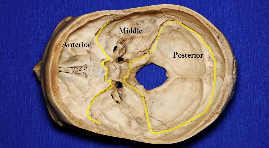
The Skull The Definitive Guide Biology Dictionary from biologydictionary.net The skull bones can be classified into two groups: During childhood development, the skull bones remain somewhat separated, allowing for growth of the brain and skull. It supports and protects the face and the brain. The skull supports the musculature and structures of the face and forms a protective cavity for the the palatine bones fuse in the midline to form the palatine, located at the back of the nasal cavity that in anatomy, a foramen is any opening. The bone is pierced by a large oval hole(the foramen magnum) through which runs the spinal cord. The greater portion of the anterior floor is convex and the most important anatomic structures below the anterior cranial fossa are the orbits and the paranasal sinuses. The skull includes the upper jaw and the cranium. A thorough description is beyond the.
These joints fuse together in adulthood.
Learn about skull base anatomy with free interactive flashcards. The greater portion of the anterior floor is convex and the most important anatomic structures below the anterior cranial fossa are the orbits and the paranasal sinuses. It is comprised of many bones, formed by intramembranous ossification, which are joined together by sutures (fibrous joints). The major sutures are the coronal suture, sagittal suture, lambdoid suture and squamosal sutures. The skull begins to form prior to week 12 of embryogenesis. Cranium) is the skeleton of the head composed of 22 separate bones joined together primarily by sutures. They don't move and united into a single unit. The bbc is not responsible for the content of external websites. The skull has a single occipital condyle.7 the skull consists of five major bones: It offers protection to the brain, eye balls, inner ears, and nasal passages. Overview, anterior skull base, middle skull base march 18, 2017. The axial & appendicular skeleton. The skull or known as the cranium in the medical world is a bone structure of the head.
The skull begins to form prior to week 12 of embryogenesis. The cranium and the mandible. These joints fuse together in adulthood. The bone is pierced by a large oval hole(the foramen magnum) through which runs the spinal cord. In order to be light, the skull is made up by flat and irregular bones, and has hollow spaces called the sinuses.
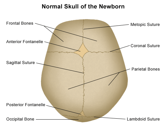
Anatomy Of The Newborn Skull from api.kramesstaywell.com Cranium) is the skeleton of the head composed of 22 separate bones joined together primarily by sutures. They don't move and united into a single unit. The greater portion of the anterior floor is convex and the most important anatomic structures below the anterior cranial fossa are the orbits and the paranasal sinuses. Inferior view of base of the skull. This anatomic region is complex and poses surgical challenges for otolaryngologists and neurosurgeons alike. Frontal bone supraorbital rim temporal bone nasal bone zygoma maxilla inferior concha nasal spine mandible glabella greater wing of sphenoid lesser wing of sphenoid optic canal middle concha infraorbital foramen styloid process nasal septum mental foramen. Upon reaching maturity, our skull bones fuse to produce a rigid protective shell for the soft nervous. A cartilaginous mould begins to grow this is why raising your eyebrows can create the appearance that the back of the head is moving.
Human anatomy for muscle, reproductive, and skeleton.
Learn more about the anatomy and function of the skull in humans and other vertebrates. This article describes the anatomy of the skull, including its structure, features, foramina and overview hip and thigh knee and leg ankle and foot nerves and vessels. Overview, anterior skull base, middle skull base march 18, 2017. Learn about skull base anatomy with free interactive flashcards. The skull is the bony skeleton of the head. Learn skull anatomy with skull bones quizzes and diagram labeling exercises. It is comprised of many bones, formed by intramembranous ossification, which are joined together by sutures (fibrous joints). Axial muscles of the head, neck, and back. The frontal, parietal, temporal and occipital bones are joined at the cranial sutures. The skull bones can be classified into two groups: The major sutures are the coronal suture, sagittal suture, lambdoid suture and squamosal sutures. A thorough description is beyond the. The bone is pierced by a large oval hole(the foramen magnum) through which runs the spinal cord.
Axial muscles of the head, neck, and back. Anatomy next provides anatomy learning tools for students and teachers. Human anatomy for muscle, reproductive, and skeleton. These joints fuse together in adulthood. Learn more about the anatomy and function of the skull in humans and other vertebrates.
Antique Engraving Illustration Clip Art Of Human Skull Anatomy Stock Photo Picture And Royalty Free Image Image 118470631 from previews.123rf.com A thorough description is beyond the. Skull bones aren't fused together at birth. Excluding ear ossicles, it is made of 22 bones. It supports and protects the face and the brain. Skull reshaping is done on any of the structures that lie above the face. The skull has a single occipital condyle.7 the skull consists of five major bones: The bbc is not responsible for the content of external websites. The base of the skull (or skull base) forms the floor of the cranial cavity and separates the brain from the structures of the neck and face.
It offers protection to the brain, eye balls, inner ears, and nasal passages.
The anterior fossa is formed by the orbital plates of the frontal bone, cribriform plate of the ethmoid, and lesser wings of the sphenoid. « back show on map ». The frontal, parietal, temporal and occipital bones are joined at the cranial sutures. The major sutures are the coronal suture, sagittal suture, lambdoid suture and squamosal sutures. The skull bones can be classified into two groups: William is a final year medical student in australia who has taught anatomy to tertiary science and. It supports and protects the face and the brain. Human anatomy for muscle, reproductive, and skeleton. Excluding ear ossicles, it is made of 22 bones. The cranium and the mandible. The occipital bone forms the back of the skull and the base of the cranium. The skull or known as the cranium in the medical world is a bone structure of the head. Anatomy next provides anatomy learning tools for students and teachers.


0 Comments